The largest organ covering the whole body
Functions of skin
Barrier against: -U.V rays (by melanin)
-Infection, trauma, dehydration (by keratin) 2-Receives sensations (by receptors)
3-Regulate body temperature (sweat glands)
1-Protection:
4-Excrete
5-Synthesis of vitamin D
6-Medico-legal importance (Finger prints) 7-Diagnosis of some diseases
(anemia, measles, chicken pox, jaundice).
waste products (sweat glands)
Types of skin
■1-Thick skin ■2-Thin skin
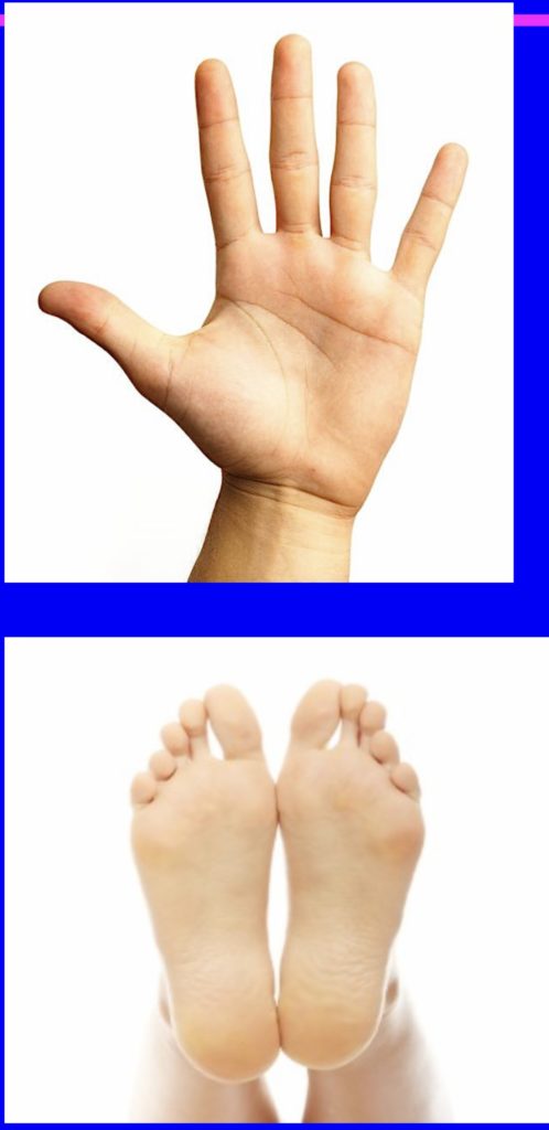
Thick Skin
■ Site:
-Palm of hands
-Sole of feet
-Tips of fingers & toes
Structure of Skin
■ ■ ■
Epidermis Dermis Skin
Appendages:
A-Hairs B-Glands C-Nails
I- Epidermis
■ keratinized stratified squamous epith.:
A-Keratinocytes:
85% of cells &
Form keratin B-Non-keratinocytes:
1-Melanocytes 2-Merkel’s cells 3-Langerhans cells
A- Keratinocytes
■5 Layers:
■1-Basal [Germinal] cell layer ■2-Spinous [Prickle] cell layer ■3-Granular layer
■4-Clear layer
■5-Horny layer
1-Basal [Germinal] cell layer
■ L.M:
1 layer of columnar cells
■
■ Resting on clear & wavy B.M
■ Basal oval nuclei [++ mitotic figures for renewal]
■ Renewal occurs every 2-4 weeks mostly at night
■ Basophilic cytoplasm
1-Basal [Germinal] cell layer
■ E.M:
■ Cells joined together by desmosomes
& to B.M by hemi-desmosomes ■ Rich in ribosomes & polysomes N.B: Melanocytes + Merkel’s cells
are present in this layer
2-Spinous [Prickle] cell layer
■ L.M:
■ 4-9 layers of polyhderal cells
■ Central rounded nuclei
■ Less basophilic cytoplasm
■ Cells have spine- like processes [Indicate sites of desmosomes as cells shrink during preparation]
2-Spinous [Prickle] cell layer
2-Spinous [Prickle] cell layer
■ E.M:
■ Cells joined by desmosomes have interdigitating
cell membranes
■ Superficial cells contain membrane
bound (coated) lamellar granules N.B: Langerhans cells are present
in this layer
■Basal layer together + Spinous layer are called Malpighian layer
3-Granular layer
■ L.M:
■ 2-4 layers of spindle- shaped cells ■ Flat nuclei
■ Basophilic granules
■ E.M: 2 types of granules:
a) Coated lamellar granules b) Keratohyaline granules
a) Coated lamellar granules
■ Oval or rod-shaped
■ Covered by membrane ■ Contain lamellar discs
[Lipid bilayers]
■ Discharge their content to
intercellular space → Lipid sheets
→ Act as cement & barrier against bacteria
b) Keratohyaline granules:
■ Large
■ Not covered by membrane ■ Form matrix →
Bind tonofilaments → Form bundles
4-Clear layer
■ L.M:
■ 1 layer of flat cells ■ No Nuclei
■ E.M:
■ No Nucleus or organelles
■ Cells joined by remnants of desmosomes
■ Contain packed filaments of eleidin [immature keratin] embedded in matrix [Formed by keratohyaline granules]
5-Horny layer
■ L.M:
■ Many acidophilic layers or scales
of keratinized dead cells
■ E.M:
■ Dead cells
■ No nuclei or organelles
■ Joined by remnants of desmosomes
■ Filled with mature keratin filaments embedded in matrix
B-Non-Keratinocytes
1] Melanocytes
■ Origin: Ectodermal ■ L.M:
■ Pale round nuclei
■ Long processes
to transport melanin to adjacent cells
■ Give Dopa +ve reaction [Dihydroxyphenylalanine]
Application of DOPA to skin → Dark color due to formation of melanin by tyrosinaze enzyme
1] Melanocytes
■ E.M:
■ Euchromatic nucleus ■ Prominent nucleolus ■ No desmosomes
between them & keratinocytes
■ Joined to B.M by hemi-desmsosmes ■ Characters of ptn. forming cells:
[+++ Mitochondria, rER & G.A]
1] Melanocytes
■ Function:
■ 1-Form tyrosinase enz.
→ Form melanin →
Gives skin colour
■ 2-Melanin granules protect DNA from UVRs
1] Melanocytes
2- Merckel’s cells:
■ Origin: Ectodermal ■ L.M:
■ Modified basal cells
[But slightly larger]
■ Naked sensory nerve fibres end in disc like expansion under these cells
2- Merckel’s cells:
■ E.M:
■ Attached to adjacent cells
by desmosomes
■ Cytoplasm contains electron-dense granules
[So, it belongs to APUD cells]
■ Functions:
■ 1-Mechanoreceptor for (touch & pressure) ■ 2-Paracrine regulation of epidermal cells
3-Langerhans cells
■ Origin: Mesodermal
■ L.M:
■ Branched cells between
cells of spinous layer ■ Dark nucleus
■ Pale cytoplasm
3-Langerhans cells
■ E.M:
■ 1ry & 2ry lysosomes ■ Well developed G.A ■ Birbeck’s granules:
[Tennis-raquet-shaped, contain hydrolytic enzs.] ■ No desmosomes
■ No junctions with keratinocytes
■ Function:
■ 1-Phagocytic cells of skin
■ 2-Antigen presenting cells [present antigens to T- lymphocytes]
II- Dermis
■ Connective tissue under epidermis
Dermis is formed of 2 layers
■Papillary layer ■Reticular layer
Papillary layer
Reticular layer
-Thinner & superficial -Loose C.T
-More cellular
-More vascular -Receptors: •Meissner’s corpuscles
-Thicker & deeper -Dense fibrous C.T -Less cellular -Less vascular -Receptors:
•Pacinin corpuscles •Krause end bulb+ •Ruffini end organ
Thick skin
Thin skin
Sties:
-Palms & soles
-Tips of fingers & toes
The rest of body
Epidermis
Thick
Thicker Malpighian layer Thicker granular (2-4Ls) Clear L present
Thick Horny layer
Thinner Thinner Thinner (1L) Absent Thinner
Dermal papillae
Regular, Numerous & High
Few Irregular
Appendages
No hair follicles
No arrector pili muscle No Sebaceous glands Numerous sweat glands
Present Present Present
Less numerous
Skin Appendages
■ ■ ■
A-Hairs B-Glands C-Nails
Thin hairy skin
A-Hairs
■ Def:
keratinized epithelial thread inserted into epidermal sheath
Structure:
1-Shaft:
Projects above skin surface
2-Root:
Embedded in skin
3-Hair bulb:
Terminal dilatation
Both shaft & root are formed of:■Medulla: Soft keratin
■Cortex: Hard keratin [contain melanin] ■Cuticle: Hard keratin
■3-Hair bulb [Terminal dilatation]
-Invaginated by vascular C.T
→ Hair papilla
-Contains:
1-Melanocytes
2-Matrix cells for growth of hair
Hair Follicle
■ Downgrowth of epidermis around root of hair ■ Formed of 3 sheaths:
1-Inner root sheath
2-Outer root sheath
3-Connective tissue sheath
1-Inner root sheath
3 layers:
• Cuticle:
Scales rich in soft keratin
• Huxley’s layer:
2-3 layers
• Henle’s layer: 1 layer
■2-Outer root sheath
■ Downward continuation of epidermis
■ At bottom → Basal layer only
■ 3-Connective tissue sheath → C.T dermis
Colour of hair
■ Caused by melanin pigment in cortex
■ Produced by melanocytes in hair matrix
■ Grey hair: In old age melanocytes fail to produce tyrosinase
■ Yellow hair: Due to pheomelanin (yellow)
■ Baldness: Hair loss due to genetic factors & sex hormones [Mainly in males]
Arrector Pili Muscle
■ Smooth muscle
■ Origin:
Papillary layer of dermis ■ Insertion:
Sheath
■ Its contraction: (By fear or cold) → Erection of
hair & depression of skin → Goose skin
B- Glands of Skin
■ 1-Sweat Glands
■ 2-Sebaceous Glands
?
a- Eccrine sweat glands
b- Apocrine seat glands
Type
Simple coiled tubular gland
Mode of secretion
Merocrine
Apocrine
Site
-All over the body -More in thick skin
-Thin skin of axilla -Pubic & perineal areas
Acini
-Lined with 2 types of cuboidal cells -Surrounded by myoepith. cells
1-Large pale [clear] cells -Broad base & narrow apex -Pale cytoplasm (glycogen)
2-Small dark cells
-Narrow base & broad apex -Dark granular cyt. (glycoptn)
a- Eccrine sweat glands
b- Apocrine seat glands
Ducts
Spiral course in epidermis & open into
Epidermis
Hair follicle
Lined with 2 layers of cubical cells
Function
→ Clear watery sweat [H2O + NaCl + urea +
ammonia]
→ Odourless sweat → by bacteria → Offensive
odour
(Start function at puberty)
2-Sebaceous gland
■ Type:
Simple or simple branched alveolar gland
■ Mode of secretion:
Holocrine
■ Site:
Associated with hair follicles
2-Sebaceous gland
■ Acini: Lined with: 1-Basal flat stem cells
to replace
2-Central polyhedral cells
with central nuclei
& vacuolated cytoplasm (fat)
2-Sebaceous gland
■ Ducts:
-Short
-Open into upper 1/3 of
hair follicle
-Lined with stratified
squamous epith.
■ Function:
Secrete sebum (oily) to prevent cracking of skin
Sebaceous gland
C-Nails
■ Def:
Plates of keratinized epith. cells
on dorsum of distal phalanx
■ Formed of:
Nail plate + Free edge + Root
(in nail groove, covered by nail fold)
■ Proximal nail fold:
Skin fold which covers the root
■ Lateral nail fold:
2 skin folds that cover lateral sides ■ Nail bed:
■ Area under nail plate: 1-Epidermal cells:
Thin malpighian layer
2-Dermis:
Highly vascular with
no papillae
■ Nail Matrix:
Malpighian cells
lining the nail groove from which the nail grows
■ Lunula: Crescent-shaped white area at the base of nail plate